Microscopy Core Facility

The Microscopy and Histology Core Facility provides you with hands-on training and access to state-of-the-art instruments to visualise both living and fixed cells. Our complete instrument line-up can be found on OpenIRIS, our booking software.
We also offer tissue processing, embedding and sectioning, vibratome sectioning and cryo-sectioning devices, as well as cell culture facilities for sample preparation. For histology, we provide support with machines and basic protocols (e.g. H&E staining, in situ hybridisation, immunostaining).
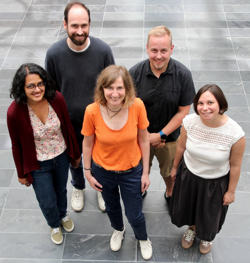
Our facility staff have a broad range of research experience and technical expertise. We are happy to provide feedback on your experimental design and assist with troubleshooting and method development at any time.
To use our services, please get in touch (microscopy(@)imb-mainz.de) to schedule a mandatory introductory session and user meeting discussing your project needs, biosafety and our user guidelines. Researchers from IMB, Mainz University, its University Medical Center and the local Max Planck Institutes are eligible to use our facility. Access for non-associated researchers is decided on a case-by-case basis.

Full service:
Full experiment design, imaging, processing, analysis and figure preparation
Self services:
- Independent booking and use of microscopes (after training)
- Independent booking and use of histology instruments (after training)
Support services:
- Comprehensive training for independent use of instruments
- Assistance with planning and designing experiments
- First level support and ad hoc troubleshooting during working hours
- Assistance with image analysis
- Proofreading of methods sections for publications
Key instruments
For a list of all our microscopes and processing stations, please click here.
Lectures and courses
We offer advanced training through practical courses and a variety of lectures. Practical courses focus on image processing and analysis (e.g. from basics to simple macro programming in Fiji, Icy, deconvolution with Huygens, Imaris) and super-resolution microscopy.
Microscopy lectures
- Introduction to microscopy
- F-techniques and super-resolution
- Histology and fluorescent labelling
- Image manipulation: the slippery slope to misconduct
Microscopy courses
Image processing and analysis:
- Basics of image analysis with Fiji/ImageJ, including filtering, segmentation, analysing objects, quantification, co-localisation, setting scale bars, visualisation of multidimensions, tracking
- 3D visualisation and analysis with Imaris and Ilastik (machine learning)
- Deconvolution with Huygens Remote Manager (HRM)
- Recording and writing macros in Fiji
- Ethics of image processing
Super-resolution microscopy:
- Introduces techniques to resolve fluorescent structures below the diffraction limit of light (ca. 250 nm in xy or 500 nm in z)
- Theoretical background of current techniques, sample preparation, hands-on STED (Stimulated Emission Depletion) and SMLM (Single Molecule Localization Microscopy), as well as data analysis

Acknowledgements/Co-authorships
“An acknowledgement of the Core Facilities in your publication is important and more than just a nice way to say thank you. Your acknowledgement matters!”
Acknowledgements are the currency by which Core Facilities are measured: they help us prove our value and our contribution to research results. Acknowledging Core Facilities shows that we are important partners in the scientific community, and it allows us to maintain funding and make further investment in the Core Facilities. In the case that Core Facility staff members have contributed significantly to your research project, we ask that you treat them the same way you would treat any other collaborator and consider a co-authorship.
In accordance with DFG recommendations, we furthermore ask that you acknowledge any instrumentation that is funded through a DFG major instrumentation grant by including the corresponding project number.
Communities
We are a member of German BioImaging (GerBI)

If you would like to get in touch with the local microscopy community alias Mainz Microscopy Connection (MMC), don´t hesitate to contact us or the MMC directly (mmc(@)imb.de).

Think global (https://microscopydb.io/who-we-are/#partners), image local.

Sandra Ritz is a principal investigator and Anusha Bargavi Gopalan is a member of the SFB 1551 on “Polymer Concepts in Cellular Function”.


